

 au
au

Brain abscesses are focal infections that are characterised by a collection of pus in the brain tissue. Opportunistic pathogens that inhabit the skin flora can gain access to the brain via head trauma or neurosurgery and cause a brain abscess. Klebsiella, Salmonella and Clostridium species are common causative agents. Dental infections can also be a cause with Prevotella spp., Bacteroides spp., and Fusobacterium spp., among the most isolated microorganisms in patients with brain abscesses [1]. Most brain and odontogenic infections are polymicrobial, so isolation of each bacterial strain present is vital for successful treatment and recovery [3]. Unfortunately, in some patients there are predispositions for brain abscesses; hereditary haemorrhagic telangiectasia (HHT) is a genetic disorder characterised by abnormal formation of enlarged blood vessels and arteriovenous malformations in organs such as the lungs, liver and brain. In more than 20% of HHT patients, there are pulmonary arteriovenous malformations, which subsequently predisposes patients to neurological conditions, such as brain abscesses [2].
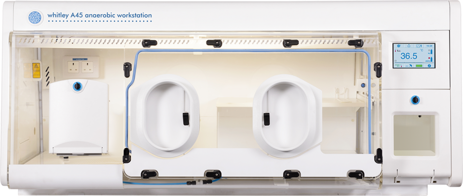
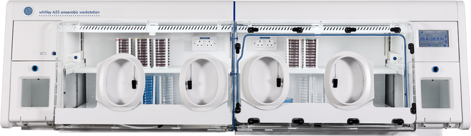
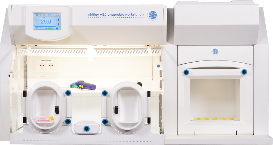
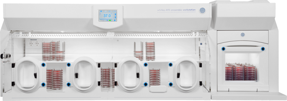
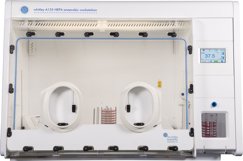
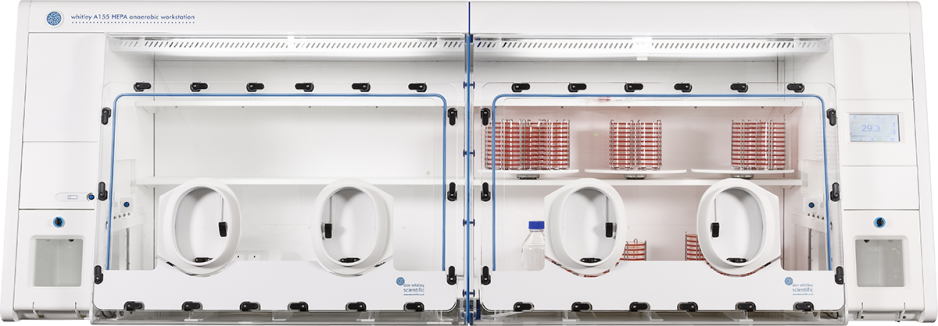
Earlier this year, a report was published detailing a case study of a HHT patient who had an odontogenic brain abscess. The patient was a 59-year-old male with HHT, who developed a brain abscess on two occasions - sadly the second occasion proved fatal. In 2012, successful treatment of a brain abscess with neurosurgery and antibiotics was first reported. The bacterial cause of this abscess was the anaerobe Fusobacterium nucleatum, a common commensal found in the human oral cavity [2]. It was suggested that poor dental health was the cause of this brain abscess [2], which is understandable, as a review from 2021 stated that around 2 – 5% of brain abscesses originated from dental infections or issues [3]. In 2022, the patient was admitted to hospital again and a CT scan showed a mass in the left parietooccipital region. Findings pointed to a brain abscess, so an acute neurological drainage was performed, and the pus collection was subject to microbiological analysis. Microscopy of the sample showed Gram-positive branching rods – it was suspected Actinomyces or Nocardia, so despite the brain abscess initially being treated with intravenous cefotaxime and metronidazole, the Gram-stain result prompted a shift to intravenous imipenem/cilastatin and trimethoprim-sulfamethoxazole. After three days, the abscess spread into the left lateral ventricle so an external ventricular drain was placed in the brain to drain fluid. The sample was cultured on agar plates both aerobically and anaerobically, and after 6 days of incubation, Arachnia propionica and Capnocytophaga ochracea was observed, so antibiotic intervention continued with the addition of intravenous clindamycin and intravenous metronidazole. The pus sample was also sent to another institution for 16S sequencing which identified three more bacterial species, Fusobacterium nucleatum, Campylobacter gracilis and Treponema medium. Minimal inhibitory concentration (MIC) testing was performed incubated in a Whitley A45 Workstation; examples of the MIC (mg/L) values was ampicillin 0.064, cefotaxime 0.125, meropenem 0.032 and benzylpenicillin 0.032. MICs were also carried out in 5% CO2 atmosphere and examples of these MICs (mg/L) were ampicillin 0.125, cefotaxime 0.032, ciprofloxacin 0.016, gentamicin 128. Further antibiotic interventions were attempted, but the patient became unresponsive to antibiotic treatment and was put on palliative care – he passed four months after the brain abscess was diagnosed [2].
This study presents novel findings of T. medium, Capnocytophaga sp. HMT-323 and Candidatus Saccharibacteria oral taxon 488 along with nine other bacteria in a brain abscess, with the cause linked to dental status [2]. Unfortunately, the infection caused proved fatal for this patient, but it is suggested that further research and understanding of these organisms in brain abscesses could aid successful outcomes and ultimately this research raises the importance of early clinical intervention.
Written by DWS Microbiologist Kirsty McTear
References
- Ruiz-Barrera MA, Santamaría-Rodríguez AF, Zorro OF. Brain abscess: A narrative review. Neurology Perspectives. 2022 July;2(3):160–7.
- Westerström P, Bivand JM, Kommedal Ø, Ehrnström B, Andreassen JS, Afset JE. Odontogenic Brain Abscess in a Hereditary Haemorrhagic Telangiectasia (HHT) Patient: Case Report with a Comprehensive Literature Review. Tropical Medicine and Infectious Disease. 2026 Mar 2;11(3):67.
- Burgos-Larraín LF, Vázquez-Portela Á, Cobo-Vázquez CM, Sáez-Alcaide LM, Sánchez-Labrador L, Meniz-García C. Brain complications from odontogenic infections: A systematic review. Journal of Stomatology, Oral and Maxillofacial Surgery [Internet]. 2022 Nov 1 [cited 2026 July 15];123(6):e794–800. Available from: https://www.sciencedirect.com/science/article/pii/S2468785522002063



 England